Thursday, November 17, 2011

Specimen #15: Mougeotia

Name: Mougeotia 
Family: Zygnemataceae
Collection Date: November 13th, 2011
Location: Southeast Pond, Hiram College Field Station, Hiram, Ohio
Collector: Katie Rumora

Key Used:

Bellinger, E. G., & Sigee, D. C. (2010). Freshwater Algae: Identification and Use as Bioindicators . Oxford : Wiley-Blackwell.

1b. Plants microscopic or if visible to the naked eye it is normally because they are present as a mass – but still requiring microscopic observation to determine the more detailed morphology … 3
3a. Cells grouped together to form a filament, strand or ribbon. Sometimes filaments can grow in such a profusion to be visible en masse, or are visible as multiseriate rows encrusting stones … 4
4a. Cell pigments localized in chloroplasts. Colour when fresh may be grass green, pale green, golden to brown, olive green or (rarely) blueish or reddish … 5
5b. Filaments or ribbons unbranched … 24
24b. Cell wall not made of silica … 30
30b. Chloroplasts not in the form of a spiral band … 31 

31b. Cells not embedded in a prominent mucilaginous envelope ... 32 
32b. Filament outline without constrictions and does not have toothed appearance ... 34
34b. Filaments many cells in length, not typically short ... 35 
35b. Alga not a large tubular thallus ... 36 
36b. Chloroplast either single or more than two per cell ... 37
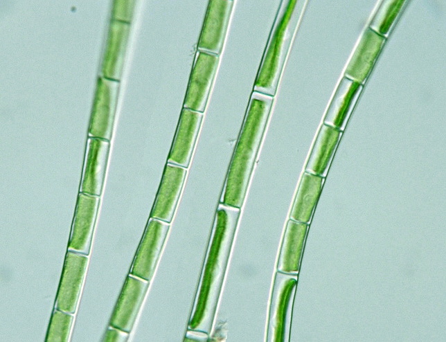
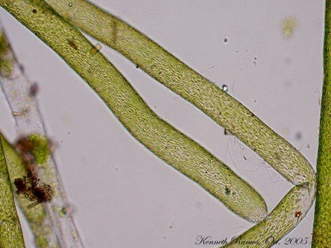
37a. One chloroplast per cell in the form of a flat plate arranged along the axis of the cell. When viewed from one direction, the chloroplast fills most of teh cell but when viewed from the other it is a thin line down the middle ... Mougeotia 


Description:
"The chloroplast of Mougeotia is suspended on cytoplasmic strands and can move within the cell depending upon the light. Hence sometimes it may be seen face-on, sometimes edge-on, and sometimes twisted. The cells form long unbranched free floating filaments. Common in many habitats" (Bellinger & Sigee, 2010).


Specimen #14 Vaucheria

Name: Vaucheria 
Family: Vaucheriaceae
Collection Date: November 17th, 2011
Location: East Hall, Hiram College, Hiram, Ohio 
Collector: Katie Rumora 

Key Used:
Bellinger, E. G., & Sigee, D. C. (2010). Freshwater Algae: Identification and Use as Bioindicators . Oxford : Wiley-Blackwell.

1b. Plants microscopic or if visible to the naked eye it is normally because they are present as a mass – but still requiring microscopic observation to determine the more detailed morphology … 3
3a. Cells grouped together to form a filament, strand or ribbon. Sometimes filaments can grow in such a profusion to be visible en masse, or are visible as multiseriate rows encrusting stones … 4
4a. Cell pigments localized in chloroplasts. Colour when fresh may be grass green, pale green, golden to brown, olive green or blueish or reddish … 5
5a. Filaments branched, sometimes rarely. False or true branching … 6
6b. Branches of filaments do not rejoin to form a net … 7
7b. Cells not in flask-shaped loricas … 8
8a. Filaments without cross walls dividing them into separate cells, cross walls only appearing when reproductive structures produced. Irregularly branched … Vaucheria

Description:
“Filaments are cylindrical, multinucleate and lack cross walls. There are numerous discshaped to oval chloroplast with or without pyrenoids. The usual storage products are oil and fat, very rarely starch. Branching does occur but its irregular. Filament 20-140µm wide. The various species are identified mainly by their reproductive structures. Widespread and may form green mats of tangles filaments in shallow freshwaters, on stones and damp soil. It may also occur in salt marshes and brackish water muds” (Bellinger & Sigee, 2010)

Figure 1: Prepared slide of Vaucheria 

Figure 2: Another view of Vaucheria

Links: 

Specimen #13: Nitzschia palea

Name: Nitzschia palea
Family: Bacillariaceae
Collection Date: November 11, 2011 
Location: Hiram College Botany Lab, Algal Tanks 
Collector: Katie Rumora 

Key Used:
Bellinger, E. G., & Sigee, D. C. (2010). Freshwater Algae: Identification and Use as Bioindicators . Oxford : Wiley-Blackwell.

1b. Plants microscopic or if visible to the naked eye it is normally because they are present as a mass – but still requiring microscopic observation to determine more detailed morphology … 3
3b. Cells individual or in groups that may be regular or irregular in shape but not forming a filament, strand or ribbon … 56
56b. Cell pigments localized in chloroplasts … 68
68b. Cells either individual, in pairs or in aggregations with no definite shape … 106
106b. Cells without flagella, non-motile … 126
126a. Cells isolated or in groups, wall siliceous with grooves or dots which form a definite pattern or other definite markings on the surface. Storage products mainly lipids and droplets may be visible within the cell. Chloroplasts one to many, yellow-green or golden to brown in colour … 127
127b. Cells elongate, cigar-, boat-, crescent-shaped, or a distorted version of these. Decorations arranged bilaterally, although this is not always obvious in cells having a crescent or distorted shape … 139
139b. Valves without long spines … 144
144b. Cells not as above … 145
145b. Cells without either costae or septa … 147
147b. Cells not triradiate … 148
148b. Cells not S-shaped in outline … 151
151b. Cells not saddle-shaped in one view ... 152
152b. Cells more narrowly elliptical, oval or elongate. Heteropolar or isopolar. One or more chloroplasts, not C-shaped … 153
153b. Cells without thickened ridges along most of the length of the raphe … 154
154b. Cells without undulate surface. Lanceolate to oval in shape, sometimes egg-shaped or narrowing towards the centre … 155
155b. Cells not as above ... 157
157b. Cells not crescent-shaped or as above … 165
165b. Cells not forming star-shaped colonies … 166
166b. Cells not as above … 167
167b. Cells not as above, isopolar and not shaped like an Egyptian mummy … 170
170b. Cells isopolar, lanceolate to elongate-elliptical in valve view, bent or curved in girdle view … 176
176b. Cells mostly solitary … 191
191b. Cells not attached, broader in shape. True raphe present … 192
192a. Two chloroplasts per cell, one either side of central axis. There are a series of carinal dots visible along one margin … Nitzschia

Description:
“Nitzchia is a large genus whose cells may be elliptical, narrow linear, spindle-shaped or sigmoid in valve view. In some species the valve centre may be slightly constricted. The raphe is displaced to one margin but the raphe on each valve is diagonally opposite the other. The raphe structure itself is a canal supported by bars which appear as carinal dots. There is no clear central area in the striate which may be fine or coarse in appearance. Two large chloroplast are present, one either end of the central area. Cells are solitary and may be benthic or planktonic. A very common and widespread genus found in a variety of water types” (Bellinger & Sigee, 2010)

Figure 1: Actual specimen of N. palea 

Links: 

Specimen #12 Hydrodictyon

Name: Hydrodictyon 
Family: Hydrodictyaceae
Collection Date: November 11, 2011
Location: Hiram College Botany Lab, Algae Tanks
Collector: Katie Rumora


Key Used:
Bellinger, E. G., & Sigee, D. C. (2010). Freshwater Algae: Identification and Use as Bioindicators . Oxford : Wiley-Blackwell.

1b. Plants microscopic or if visible to the naked eye it is normally because they are present as a mass – but still requiring microscopic observation to determine the more detailed morphology … 3
3a. Cells grouped together to form a filament, strand or ribbon. Sometimes filaments can grow in such a profusion to be visible en masse, or are visible as multiseriate rows encrusting stones … 4
4a. Cell pigments localized in chloroplasts. Colour when fresh may be grass green, pale green, golden to brown, olive green or (rarely) blueish or reddish … 5
5a. Filaments branched, sometimes rarely. False or true branching … 6
6a. Branches of filaments rejoin to form a net ... Hydrodictyon

Description:
“Hydrodictyon (commonly known as the ‘water net’) forms macroscopic nets that are free floating (occasionally attached) of many centimeters in length. Populations may be large enough to partially block small streams, espically nutrient-rich waters. The cells are cylindrical and joined at the ends to form a net. Individual adult cells can be several millimeters in length … Widespread in ponds and slow moving ditches” (Bellinger & Sigee, 2010)

Figure 1: Prepared slide of Hydrodictyon 

Figure 2: Actual Specimen of Hydrodictyon 

Links: 

Specimen #11: Giminella

Name: Giminella
Family: Gleoutilaceae
Collection Date: November 15th, 2011
Location: Hiram College Botany Lab Algal Tanks
Collector: Katie Rumora


Key Used:
Bellinger, E. G., & Sigee, D. C. (2010). Freshwater Algae: Identification and Use as Bioindicators . Oxford : Wiley-Blackwell.
1b. Plants microscopic or if visible to the naked eye it is normally because they are present as a mass – but still requiring microscopic observation to determine the more detailed morphology … 3
3a. Cells grouped together to form a filament, strand or ribbon. Sometimes filaments can grow in such a profusion to be visible en masse, or are visible as multiseriate rows encrusting stones … 4
4a. Cell pigments localized in chloroplasts. Colour when fresh may be grass green, pale green, golden to brown, olive green or (rarely) blueish or reddish … 5
5b. Filaments or ribbons unbranched … 24
24b. Cell wall not made of silica … 30
30b. Chloroplasts not in the form of a spiral band … 31
31a. Filaments are unbranched with the cells embedded in a prominent broad mucilaginous hyaline cylindrical envelope with broadly rounded ends… Geminella

Description:
“Cells are between 5-25µ broad and up to twice as long as they are wide and are surrounded by a thick sheath of mucilage. Cells often occur in pairs along the filament. The chloroplast is a parietal plate, roughly saddle-shaped and located near the central region of the cell. A pyrenoid is usually present. Reproduction is by fragmentation of the filament. Occurs in shallow, often slightly acidic, waters. Chlorophyta” (Bellinger & Sigee, 2010).

Figure 1: Prepared slide image of Giminella 

Figure 2: Actual specimen of Giminella 

Links: 


Thursday, November 10, 2011

Specimen #10: Cladophora

Name: Cladophora
Family: Cladophoraceae
Collection Date: November 6th, 2011
Location: Presque Isle, Pennsylvania
Collector: Willa Schrlau

Key Used:
Bellinger, E. G., & Sigee, D. C. (2010). Freshwater Algae: Identification and Use as Bioindicators . West Sussex: Wiley-Blackwell.

1b. Plants microscopic or if visible to the naked eye it is normally because they are present as a mass – but still requiring microscopic observation to determine the more detailed morphology … 3
3a. Cells grouped together to form a filament, strand or ribbon. Sometimes filaments can grow in such profusion to be visible en masse, or are visible as multiseriate rows encrusting stones … 4
4a. Cell pigments localized in chloroplasts. Colour when fresh may be grass green, pale green, golden to brown, olive green or (rarely) bluish or reddish … 5
5a. Filaments branches, sometimes rarely. False or true branching. Filaments may branch only occassionaly so it is important to examine a reasonable length to determine whether they branch or not … 6
6b. Branches of filament do not rejoin to form a net … 7
7b. Cells not in a flask-shaped loricas … 8
8b. Filaments with normal cross walls, not siphonaceous …
9b. Filaments not multiseriate … 13
13b. No hairs or setae present … 18
18b. Branched filaments not embedded in mucilage ... 21
21b. Plants larger. Cells greater than 7µm in diameter. The first cross walls of each branch occurs at the origin of the branch. Pyrenoids are present … 22 
22b. Filaments tapering abruptly, not gradually, with a rounded blunt end cell … 23
23b. Branched often longer and more robust. Repeated branching may occur … Cladophora

 
Figure 1: Cladophora


Figure 2: One view of a preserved specimen of Cladophora 


Figure 3: Another view of a preserved specimen of Cladophora 

Description:
“Cladophora is typically well branched but in gently flowing waters branching may be intermittent and difficult to find. In contrast, on a lake shoreline habitat subject to choppy wave action, the form may be tufted or bushlike with many branched. Cladophora may be free living or attached to a substrate by means of a small holdfast. The branches may be alternate or opposite, dichotomous or even trichotomous. Cell walls are robust and the chloroplast net-like (reticulate) and parietal with numerous pyrenoids. Cells 50-150µ broad and up to 10x long as broad. Commonly known as “blanket weed” it can form extensive, coarse to touch mats. Frequent in hard or semi-hard waters especially those enriched with sewage. May also be found in marine habitats especially those enriched with sewage. Cladophora can also produce large growths in water treatment filtration systems such as slow sand filters where severe problems of filter clogging may result” (Bellinger & Sigee, 2010).


Links:

References:
Bellinger, E. G., & Sigee, D. C. (2010). Freshwater Algae: Identification and Use as Bioindicators . West Sussex: Wiley-Blackwell.